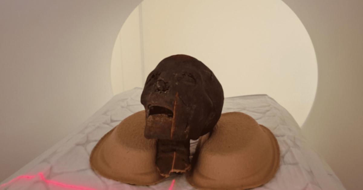
 Researchers in Hungary are uncovering remarkable new details about ancient Egyptian mummies using one of the world’s most advanced medical imaging technologies.
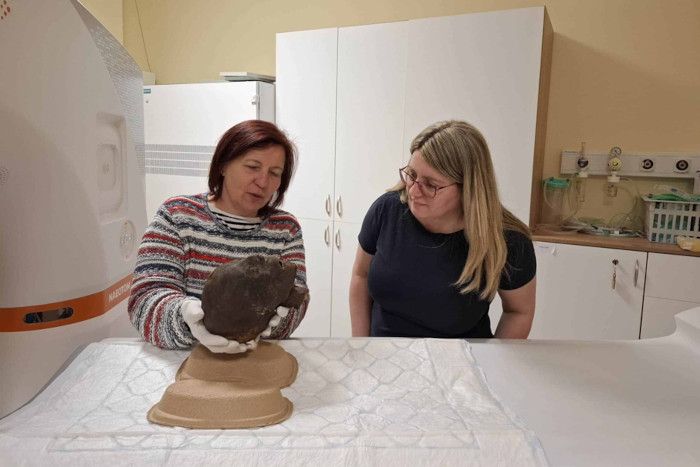
At Semmelweis University, scientists have begun examining centuries-old mummified remains with a cutting-edge photon-counting CT scanner capable of producing extraordinarily detailed internal images without damaging the fragile artifacts.
The project, conducted in collaboration with the Hungarian National Museum and the Semmelweis Museum of Medical History, is already revealing information that older scanning technology simply could not detect.
Researchers say the new images are helping them better understand ancient diseases, burial customs, mummification techniques, and even the identities of individuals preserved for more than 2,300 years.
Seeing Inside Ancient Mummies Like Never Before
The examinations focused on several Egyptian remains that have been housed in museum collections for decades, including mummified heads, limbs, and partially preserved body fragments.
Although some of these artifacts had previously undergone conventional CT scans, earlier technology lacked the resolution necessary to fully analyze the dense layers of bone, textiles, embalming materials, and preserved tissue.
The new photon-counting CT system changes that dramatically.
Unlike traditional scanners, photon-counting technology measures individual X-ray photons in far greater detail, producing sharper images with improved contrast and significantly reduced visual noise.
According to Dr. Ibolyka Dudás, head of the post-mortem imaging working group at Semmelweis University, the goal is to create the clearest possible picture of the internal structures, preservation methods, and any medical abnormalities present within the remains.
Detailed Insights Into Ancient Lives
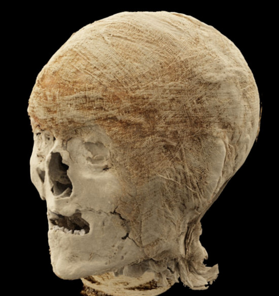
Among the most important specimens examined were two mummified heads.
The high-resolution scans allowed researchers to closely study dental structures and skull sutures with a level of clarity never previously achieved. These details may help scientists estimate the individuals’ ages more accurately and potentially create advanced 3D facial reconstructions in the future.
Researchers also examined a mummified lower leg that had long puzzled scientists.
The new scans suggest the individual may have suffered from osteoporosis, although further study will be required to determine whether the condition resulted from aging or disease.
A second preserved limb appears to belong to a young individual, providing researchers with the first highly detailed anatomical data ever collected from the specimen.
A “Mummy Bundle” Revealed To Be Something Else Entirely
One of the most surprising findings involved a mysterious wrapped object that had originally been classified decades ago as either a human head or possibly even a bird mummy.
Earlier scans hinted that this assumption was incorrect. The new imaging technology has now confirmed that the object is actually an adult human foot.
Researchers say the scans clearly reveal multiple layers of wrapping textiles and their differing structural characteristics, which may provide important clues about ancient mummification techniques and burial preparation.
The remains were likely once part of a complete mummy, though experts still do not know when or why the body was separated.
 Unlocking Ancient Health Mysteries
Another specimen currently under examination is a mummified hand.
By studying bone size, development, and morphology, researchers hope to determine whether the remains belonged to a child or an adult and possibly identify the individual’s biological sex and approximate age.
Radiocarbon dating has already established that some of the examined remains date between 401 and 259 BCE, placing them more than 2,300 years in the past.
Scientists believe the ongoing research could provide valuable information about ancient Egyptian health, disease, nutrition, injuries, and funerary practices.
A New Era for Mummy Research
Experts say the project demonstrates how rapidly advancing imaging technology is transforming archaeology and bioanthropology.
Because the scans are completely non-invasive, researchers can study delicate remains in extraordinary detail without physically opening or damaging the mummies themselves.
Chief Museologist Krisztina Scheffer noted that the technology is already revealing information hidden inside the remains for thousands of years.
For archaeologists and historians, the work at Semmelweis University represents a major step forward in understanding not only how ancient Egyptians prepared their dead, but also how they lived.
As image analysis continues, researchers expect even more discoveries to emerge from the scans — proving that some of history’s oldest secrets are still waiting to be uncovered beneath layers of linen and time.